


Brian Choi, Bio-application scientist
For more information, please contact 该Email地址已收到反垃圾邮件插件保护。要显示它您需要在浏览器中启用JavaScript。
Data Reference: Prof. Ushiki and Dr. Nakajima, Niigata Univ.
The SICM image shown below is the world’s first observation of rat tracheal tissue in an aqueous environment. The luminal surface of the tracheal tissue was successfully imaged using Park SICM. For comparison, the same position of the tissue was imaged by scanning electron microscopy (SEM) after SICM measurement. To avoid structural damage to the sample, the tissue was very carefully dehydrated by a highly skilled researcher for SEM sample preparation. Highly ciliated cells are scattered on the luminal surface of the tracheal tissue. The hair-like cilia are easily waved by liquid flow in nature.
- Directly acquire physiological tissue images or single-cell images in an aqueous environment, without a laborious sample preparation procedure, with high resolution equal to SEM
- Cilliated cell in tracheal tissue imaging, not losing a single hair-like cilium in physiological conditions
Although the SICM pipette must approach this delicate surface repeatedly, every cilium in the imaged cell is visualized accurately, in contrast to the SEM image. Both ciliated and non-ciliated cells of tracheal tissue are pictured, especially the hair-like details of ciliated cells. This result is proof that scientifically explains with the SICM technique that in an aqueous environment we can directly acquire physiological cell and tissue images with a resolution equal to that of SEM.
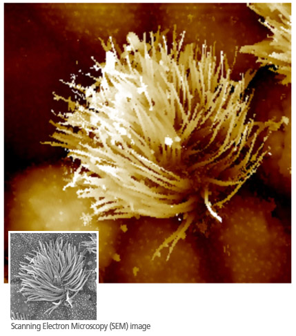 SICM, Ciliated Cell in Tracheal Tissue
 Biological Tissue Preparation for SEM Imaging
The scanning electron microscope (SEM) has been an indispensable tool for cell biology due to its high-resolution cell imaging capability. To acquire an SEM image of a cell surface, a laborious sample preparation procedure, such as freeze-drying the whole cell and metal-coating the cell membrane, is inevitable. Because of this preparation, the cells are no longer considered living, but biologically dead. Moreover, the dead sample must stay in a vacuum chamber to ensure that electron paths remain straight; thus, SEM is not applicable under liquid conditions.
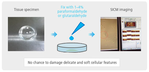 Biological Tissue Preparation for SICM Imaging
In SICM imaging, the complicated sample preparation process of SEM imaging, which often causes physical damage of cellular and sub-cellular structures, is unnecessary. SICM frees researchers from the risk of artifacts and imaging failure. This technical advantage of SICM results in experimental high productivity for research and routine imaging needs.
Park Cell Analysis Systems
|
|
|
|
| Park NX12-Bio | Park NX10 | Park XE7 | |
| Scanning Ion Conductance Microscopy (SICM) | |||
| Atomic Force Microscopy (AFM) with liquid probe hand | |||
| Inverted Optical Microscopy (IOM) | |||
| Live Cell Chamber |