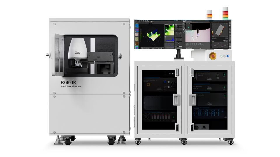
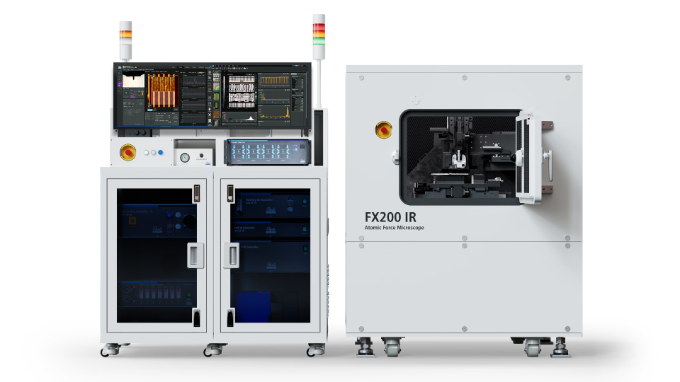
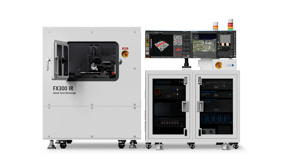
Park nano-IR spectrometers seamlessly integrate atomic force microscopy (AFM) with photo-induced force microscopy (PiFM) technology. These innovative platforms deliver molecular vibration mapping at the nanometer scale while maintaining full AFM capabilities for comprehensive topographic, nanomechanical, electrical, and thermal analysis.
With simultaneous acquisition of nano-IR chemical information and surface topography, Park nano-IR spectrometers are ideal for nanoscale IR spectroscopy and imaging in semiconductors, polymers, thin films, 2D materials, and other advanced materials.
Key Features
Streamlined Automated Workflow for Nanoscale IR Measurements
Park nano-IR spectrometers build upon the proven automation of the FX platform, including probe exchange and vision-guided AFM laser alignment, and extend it with advanced automation dedicated to IR measurements.
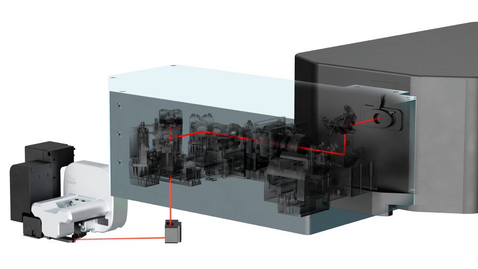
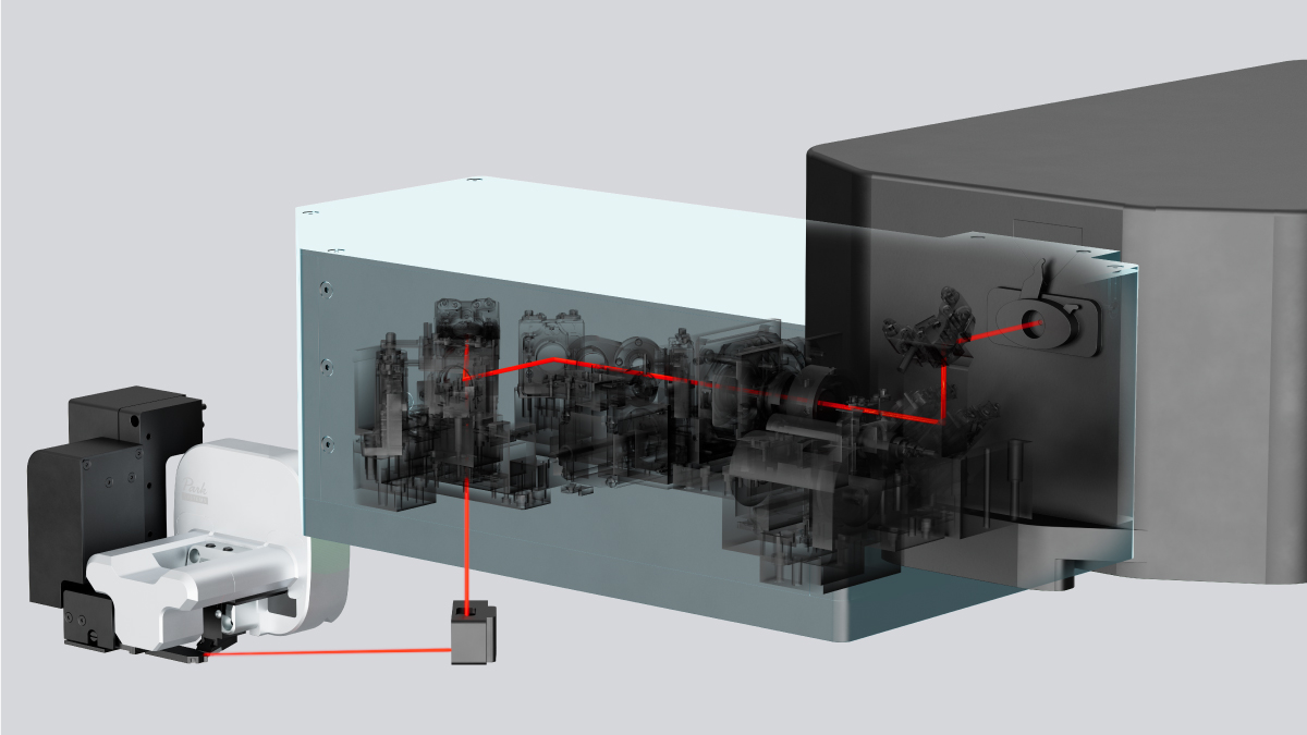
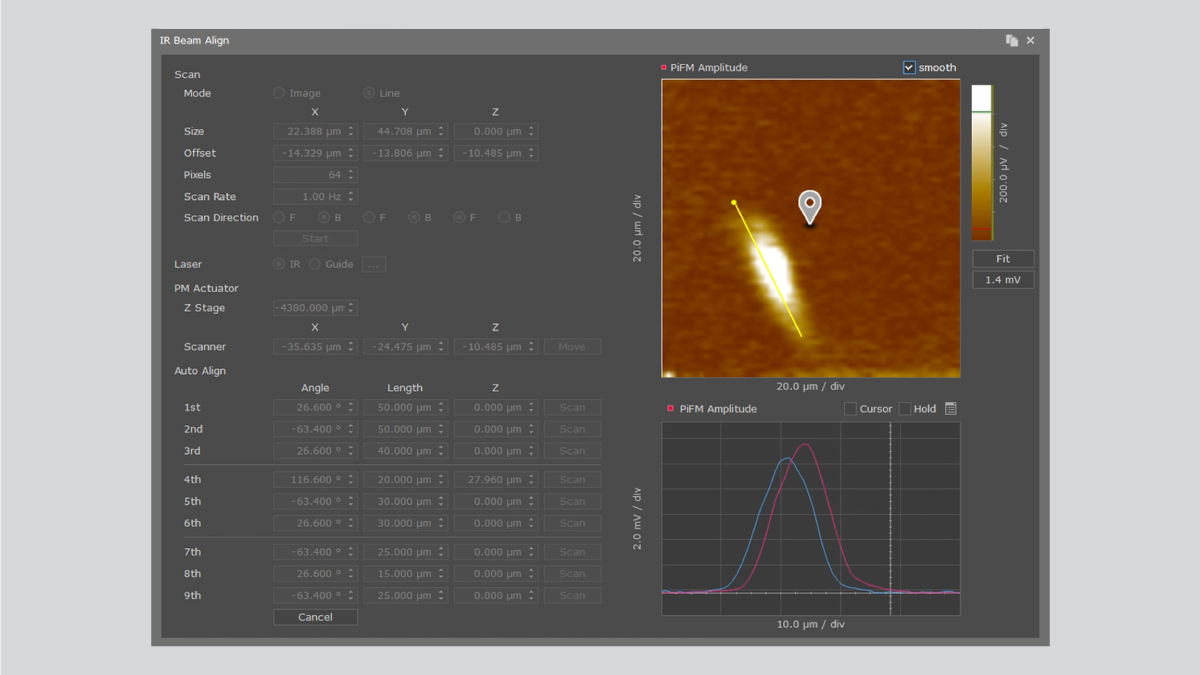
In particular, the system features a fully automated IR beam alignment routine that uses the measured nano-IR signal as real-time feedback to locate the optimal beam position.
Automatic IR Beam Alignment Powered by SmartScan™
SmartScan™ performs automatic IR beam alignment. After executing a full-stroke scan using the parabolic mirror (PM) actuator, the system automatically identifies the position where the measured nano-IR signal reaches its highest intensity. This ensures accurate alignment and maximizes signal quality with minimal user intervention.
True Non-Contact™ IR Measurements
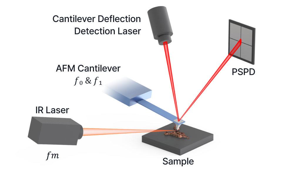
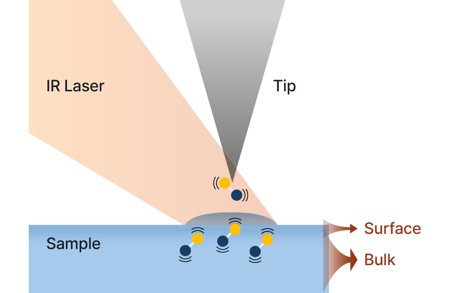
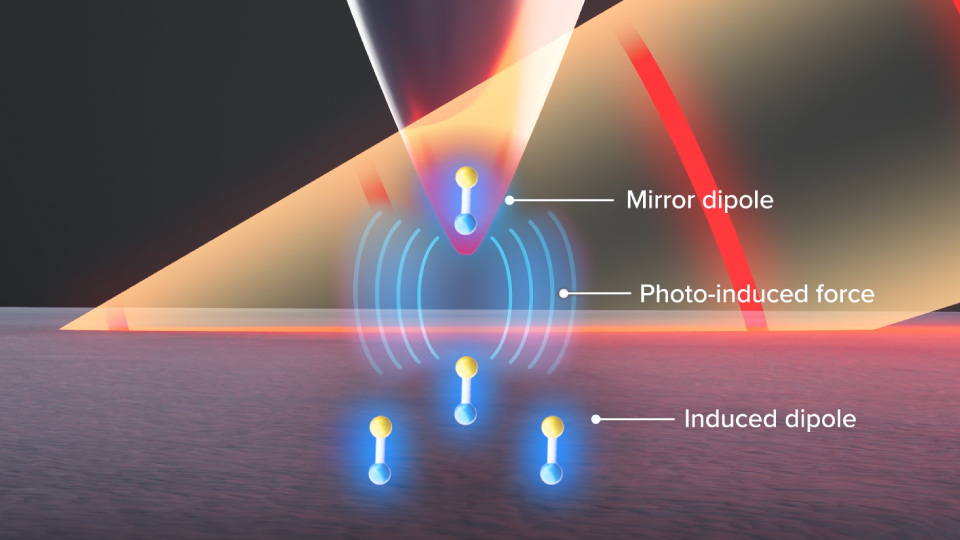
Park nano-IR spectrometers leverage Photo-induced Force Microscopy (PiFM) to perform fully non-contact infrared measurements, detecting the photo-induced force between the AFM tip and the sample without ever touching the surface. This approach eliminates sample damage and tip contamination inherent in contact-based techniques, achieving sub-5 nm spatial resolution
and extending probe lifetime.
Non-contact mode feedback maintains precise tip–sample separation
throughout the measurement, ensuring high reproducibility on soft or
delicate materials. With streamlined operation, users can focus on results rather than setup, making nano-IR analysis more reliable and accessible than ever.
Selectable Detection Modes for Nano-IR Measurements
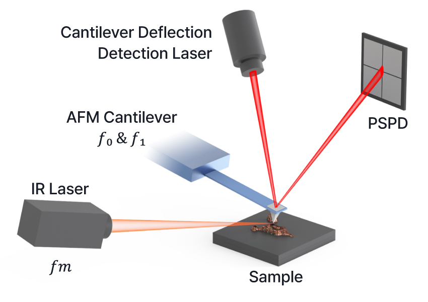
Park nano-IR spectrometers offer two distinct detection modes, enabling users to optimize measurements based on sample characteristics and analysis goals. Direct mode tunes the laser to the cantilever’s resonance frequency, maximizing signal strength and making it effective for probing broader or deeper sample responses.
Sideband mode modulates the laser at the difference frequency ( 𝑓₁ - 𝑓₀ ), selectively detecting nonlinear interactions at the tip–sample interface. This allows for highly localized chemical contrast with minimal background, ideal for resolving fine features on complex or heterogeneous surfaces.
Optimized Detection Frequency for Stable Nano-IR Imaging
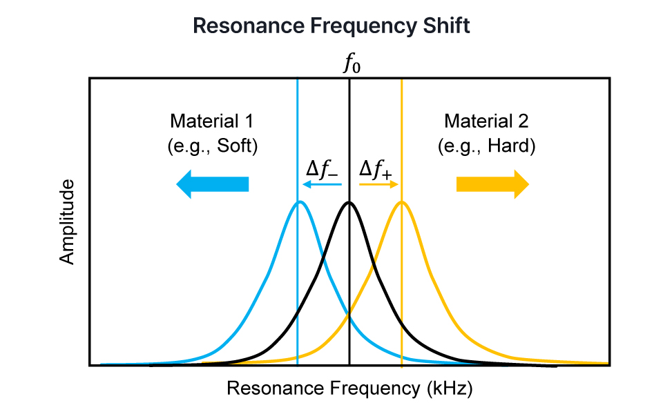
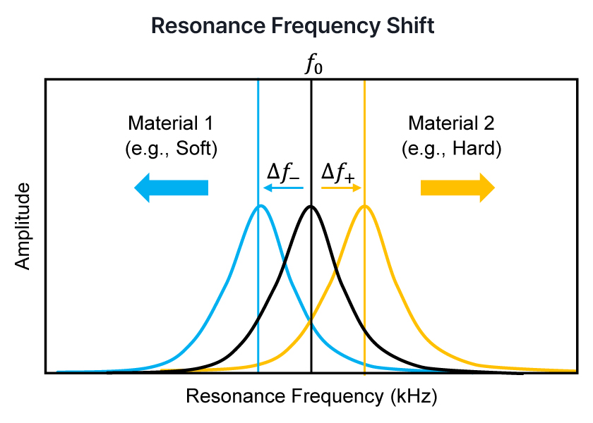
Consistent, high-resolution nano-IR imaging requires more than just resonance. It demands precise frequency tracking. As the AFM tip scans across materials with varying mechanical properties, the tip–sample interaction can shift the cantilever’s resonance frequency. If not properly adjusted, this detuning may lead to signal degradation and mechanical artifacts.
Resonance Frequency Shift Material 1 (e.g., Soft) Park nano-IR spectrometers address this by automatically scanning a narrow frequency range at every pixel and selecting the optimal detection frequency. This ensures the system stays aligned with the actual resonance condition throughout the scan, maintaining strong signals, reducing artifacts, and improving signal-to-noise ratio (SNR) for reliable chemical imaging on heterogeneous samples.
Park AFM Technology
High Resolution Chemical Imaging with Nano-IR
Photo-induced Force Microscopy (PiFM) exploits the photo-induced force generated when a tunable IR laser excites molecular vibrations at their resonant frequencies. In the Park nano-IR spectrometers, the AFM cantilever operates in Non-contact mode to detect these forces without ever touching the sample surface.
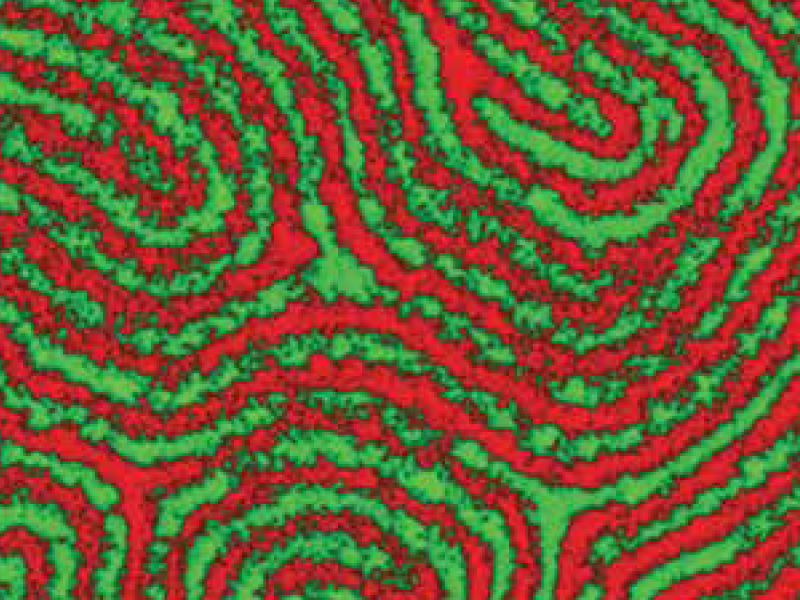
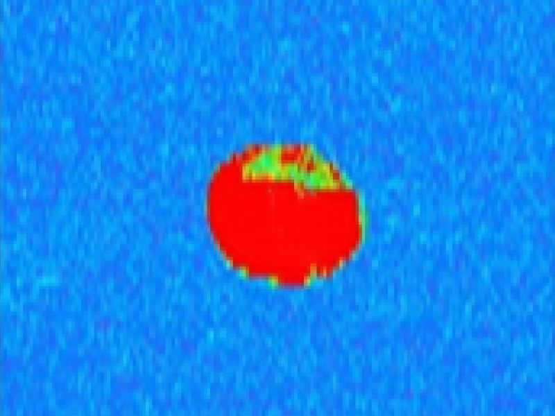
As the laser wavelength is swept, changes in cantilever oscillation amplitude directly reflect local IR absorption, producing both spectral and chemical maps. This fully non-invasive approach delivers true nanoscale chemical characterization—achieving sub-5 nm spatial resolution and monolayer sensitivity—enabling researchers to visualize molecular composition at the atomic scale.